Logo
Phone: 775-323-3000 | Fax: 775-323-3001
689 Sierra Rose Dr, Unit B | Reno, NV 89511

Office-based Interventional Suite (OIS)

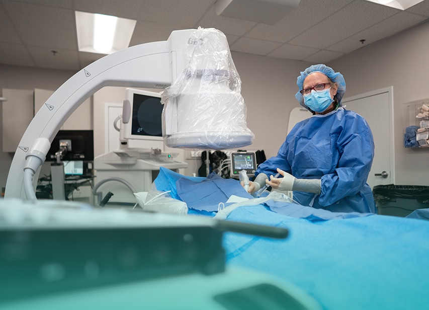
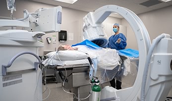
In early 2019, Nevada Vein and Vascular completed the build-out of Reno’s first Office-based Interventional Suite, or OIS. An OIS is a specialized room where minimally-invasive endovascular procedures are performed. Our state-of-the-art facility incorporates the latest in imaging technology and is fully-stocked with a large variety of tools which allow us to perform both simple and complex peripheral vascular interventions that would otherwise need to be done in the hospital.

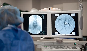
We perform a wide variety of diagnostic and therapeutic interventions in the OIS including diagnostic angiography, abdominal and lower extremity endovascular revascularizations including complex limb salvage procedures, iliac stenting, upper extremity angiography, and fistulograms with interventions for dialysis patients.

These same day procedures are completed under minimal to moderate conscious sedation. Our trained staff and physicians continuously monitor patients during the procedure and throughout their recovery to provide a safe experience and allow a quick recovery and return home with minimal to no side effects.

Why Choose Nevada Vein & Vascular for your PAD procedure?

Hospital-based procedures are expensive and time-consuming. At NVV, we understand that your time is valuable, and we’ll work with you to schedule procedures around your life. Our OIS affords our patients numerous other benefits as well. By avoiding the hospital for your procedure, you may save significantly on out-of-pocket expenses, insurance billing is reduced, check-in and wait times are decreased by many hours, and family is able to stay close by prior to and after the procedure.

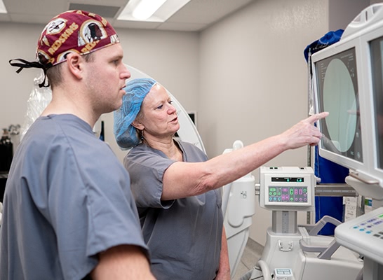
We offer the best outpatient experience possible by providing timely access to the care you need, tailored to your specific condition. Our knowledgeable staff is dedicated to our patients and every procedure is performed with meticulous attention to detail.

Vascular surgeons are the only medical specialists trained to perform both minimally-invasive and open surgical procedures. This distinction is critical when it comes to treating vascular disease because there are benefits and limitations to each approach. With this unique expertise, we sometimes combine endovascular and open surgery at the same time to perform what is known as a “hybrid” procedure. While these complex operations are performed in the hospital, hybrid procedures may reduce the number of interventions, number of necessary hospital admissions, cost, and down-time to our patients because we treat multiple areas in a single setting.

Remember that you always have a choice in where you receive vascular care. Call today 775-323-3000 to schedule your appointment with one of our vascular specialists.

Procedures we offer in the office

Please explore the content below

In medical jargon, endo simply means “inside” and “vascular” implies arteries or veins. An endovascular procedure is one which is performed within the blood vessel itself.

Typically, the femoral artery in the groin is accessed with little more than a needle stick and there is no major incision.

To navigate the vascular system, surgeons use specialized x-ray equipment and contrast dye to highlight vascular anatomy. Wires with different characteristics are inserted. Some of these wires are very slick, others are floppy, some are very stiff, and they have different shapes on the end. We also use catheters which are similar to very long straws through which we place the wires. They too have various properties and come in a large array of sizes, shapes, and stiffness.

To revascularize, or open a blockage in a blood vessel, we use a different tool kit which includes small balloons inflated within, or stents which are made of alloy metals formed into tubes to keep the artery open. A third option to treat a blockage is a catheter with a specialized tip that spins at very high RPMs to open a channel in the vessel. This is called an atherectomy, (ather-from atherosclerosis, and “ectomy” means to remove), or you may hear the term “roto-rooter.” At times, all three options are needed for revascularization.

Diagnostic Angiogram

An angiogram is a diagnostic procedure to evaluate arterial anatomy and assess the severity of any blockages. For patients with AIOD or PAD, an angiogram is required before any treatment can be performed, but it is done at the same time as angioplasty or stenting.

Contrast dye is injected into the blood stream to define the anatomy. The dye absorbs x-rays, thereby making the blood vessels appear dark. Based on the findings, we then determine the best treatment option in real-time. In some cases, there is no suitable endovascular option and open surgery is necessary. In these instances, the angiogram provides all the necessary information to plan an open surgery.

Carbon Dioxide (CO2) Angiography

CO2 angiography is reserved for patients with kidney disease. Regular contrast dye is generally very safe, but it can be damaging to the kidneys, particularly in patients with severe pre-existing renal problems. In our OIS, we offer an excellent alternative: CO2 angiography.

Rather than dye, carbon dioxide is injected as the contrast agent to define the arterial anatomy. CO2 has no ill-effects on the kidneys, yet is still very effective. If an intervention such as angioplasty, atherectomy, or stenting is required, a small amount of contrast will ultimately be needed, but the overall amount is reduced to a fraction of what would otherwise be required.

Intravascular Ultrasound (IVUS)

Intravascular ultrasound, or IVUS (eye-VUS), is an invaluable tool we use to assess blood vessel anatomy from within the vessel itself. A tiny ultrasound probe is inserted into the blood vessel to obtain high-resolution images of all three layers of the vessel wall. IVUS also allows for real-time imaging of a blockage to evaluate its severity, and for immediate assessment of the effectiveness of angioplasty or stenting.

IVUS has an added benefit in that we use it together with either contrast angiography or CO2 angiography to further reduce the total amount of contrast required for a procedure.

Angioplasty

During an angioplasty, a blocked artery is opened using an inflatable balloon. These balloons come in a variety of sizes and lengths which we match to the diameter of the artery and length of the blockage. A channel in the artery is created to restore flow. After an angioplasty, nothing is left behind in the artery. If a blockage is severe, it may be used in combination with stenting and/or atherectomy.

Iliac Stenting

The iliac arteries are found in the pelvis and supply the entire blood volume to the legs. Blockages here are frequently calcified, dense, and inelastic. Because of these characteristics, iliac stenosis is most commonly treated with stents. Whenever a stent is placed, we also perform an angioplasty with a balloon. The balloon ensures the stent is dilated sufficiently enough to maximize blood flow.

Peripheral Arterial Stenting

Peripheral arterial stenting refers to stents placed in the legs. It is an effective option to treat severe lifestyle-limiting claudication and critical limb ischemia in patients with significant blockages in the arteries. The stent is advanced through the blockage and then expanded to essentially force the artery open. Angioplasty is then performed to ensure the stent achieves the appropriate diameter.

Atherectomy

Atherectomy is a procedure in which a specialized rotating catheter is inserted into a blocked artery to actually remove atherosclerotic plaque and open a channel through which blood will flow. The tip of the catheter spins at more than 100,000 RPMs to pulverize the blockage. The catheter also has suction to remove the minute fragments. Angioplasty is then performed to help expand the vessel, and in some cases, a stent may also be used.

Venography & Venous Stenting

Patients with a history of iliofemoral DVT, venous stenosis, or May-Thurner Syndrome have a blockage in the iliac veins in the pelvis. These veins are the main drainage for the leg and if flow is compromised, severe lower extremity swelling may result. We are able to diagnose and treat this condition in the OIS with a venogram and stenting.

Venography is identical to angiography, but it is used to evaluate the veins. Once the venous anatomy and blockage is identified, a stent is frequently needed. Venous stents are identical to arterial stents, but are usually a bit larger in diameter.

Fistulogram

Patients with fistulas or AV grafts may develop problems during dialysis. If you have noticed prolonged bleeding after dialysis or low flow during dialysis, there may be a treatable problem. It is always better to treat these issues before your access stops functioning. We are pleased to offer an outpatient option for our dialysis patients in our OIS, helping you to avoid a hospital visit or admission.

A fistulogram is a procedure to diagnose and treat access problems. Contrast dye is injected to identify any blockage or narrowing. Most commonly, angioplasty is performed to improve blood flow and allow dialysis to continue. For recurrent blockages that have required several interventions, or narrowing of the large central veins in the chest, a stent may also be needed.

Once a fistula or graft develops problems, they frequently recur. Our team will ensure you have frequent follow-up for any necessary maintenance.

Frequently asked questions

Will I be exposed to radiation during a procedure?

All endovascular procedures require the use of x-ray equipment. Our OBL is equipped with a state-of-the-art GE OEC 990 Elite C-arm (can we insert a link to GE website for this??) which allows for crystal-clear imaging and minimization of radiation exposure. All medical personnel at our office who use x-ray technology receive extensive training in radiation safety and we employ the ALARA (make this a link to ALARA info) (As Low As Reasonably Achievable) principles to keep exposure to a minimum.
Additionally, we have a specialized catheter called IVUS (Intra-Vascular Ultrasound) which allows us to evaluate blood vessels from the inside without using radiation at all.

What if I’m allergic to iodine or contrast dye?

Iodine is an essential trace element that all humans need for various metabolic functions. It is present in high quantities in table salt, dairy products, shell fish, tuna, and eggs to name a few common foods. It is not possible to be allergic to iodine itself. However, iodine can form compounds with other molecules and it is possible to form an allergy to these substances.

Contrast dye is a substance that incorporates iodine in its chemical makeup. The dye blocks the passage of x-rays, thereby creating a silhouette of the blood vessels which surgeons use to diagnose and treat blockages. Contrast is generally safe for most people, but reactions to dye and allergies do occur. A common reaction when dye is injected is a feeling of warmth or flushing. This is not an allergy. True allergies are less common but include a rash, itching, swelling of the lips and/or tongue, difficulty breathing, or low blood pressure. If you have a history of these types of reactions, it is very important to let your surgeon and our staff know ahead of time.

If you are allergic to dye, we may need to give you medication including a steroid dose pack and Benadryl starting the evening before your procedure, but in most cases, interventions can be done safely.